Instruments
Renishaw inVia Confocal Raman Microscope (CRM)
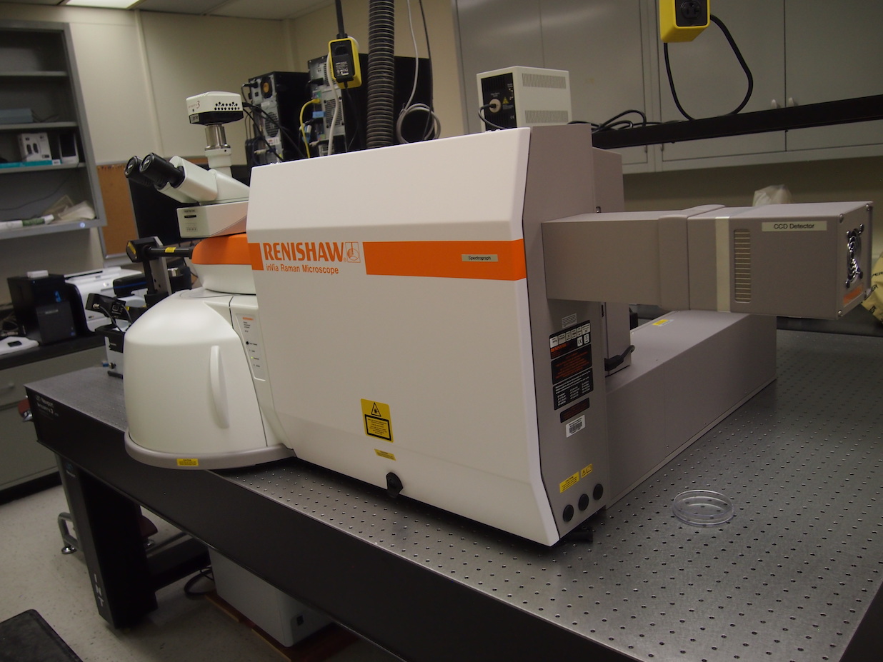
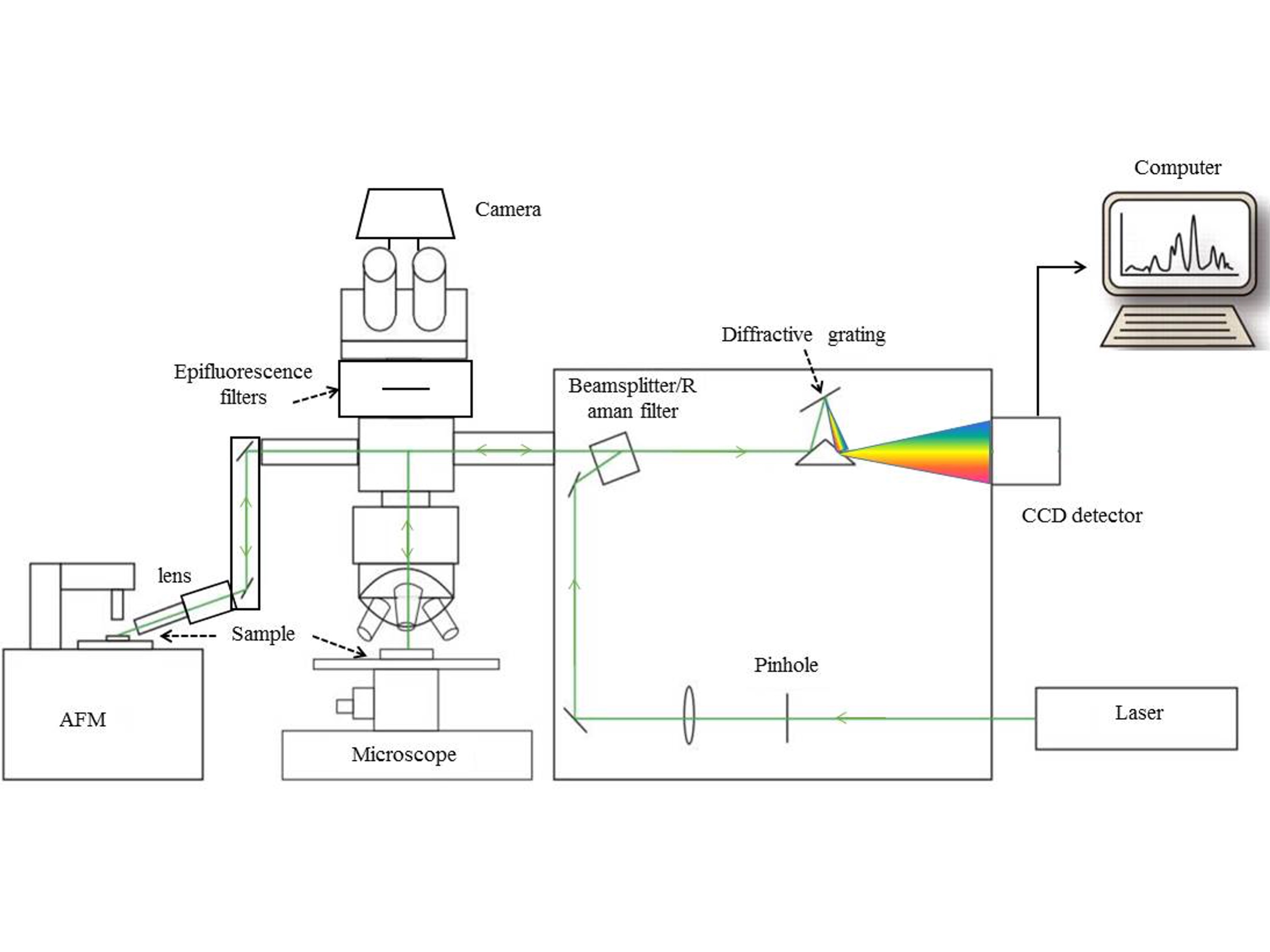
The central research instrument is an inVia High Resolution Confocal Raman Microspectrometer, which includes:
• a 633 nm He/Ne laser (17mW)
• a 457/514 nm Ar ion laser (150mW)
• a 785 nm diode laser (300mW)
• 600, 1200, and 1800 l/mm spectrographic gratings and appropriate filters with > 30% optical throughput
• a thermo-electric (TE) cooled 1024×256 CCD detector achieving a spectral resolution of 0.5 cm-1 @ 680 nm using the 1800 l/mm grating
• main sampling platform is a Leica DM2500 upright microscope with computer-controlled motorized stage for XYZ mapping with <0.1 um step-size; 10x, 20x, 40x, 50x, 50xLWD, 63x and 100x objective lenses; operates in transmitted, reflected, bright field, dark field, epifluorescent, and Raman chemometric modes; DAPI, FITC, CY3, and CY5 fluorescent filter sets available; 150 mW Hg lamp for epifluorescence
• Raman spatial resolution – diffraction limited to ~200 nm @ 457 nm and 100x magnification
• Streamline spectrograph upgrade for rapid 2D and 3D Raman scans
• Luminera Infinity 3 TE cooled camera for high-res video images and Renishaw camera for laser focusing
• Linkam THMS600 Hot/Cold stage (-196 to +600°C) with T95 controller, enabling programmable local and remote control
• video monitors, workstation and WiRE 4.1 software
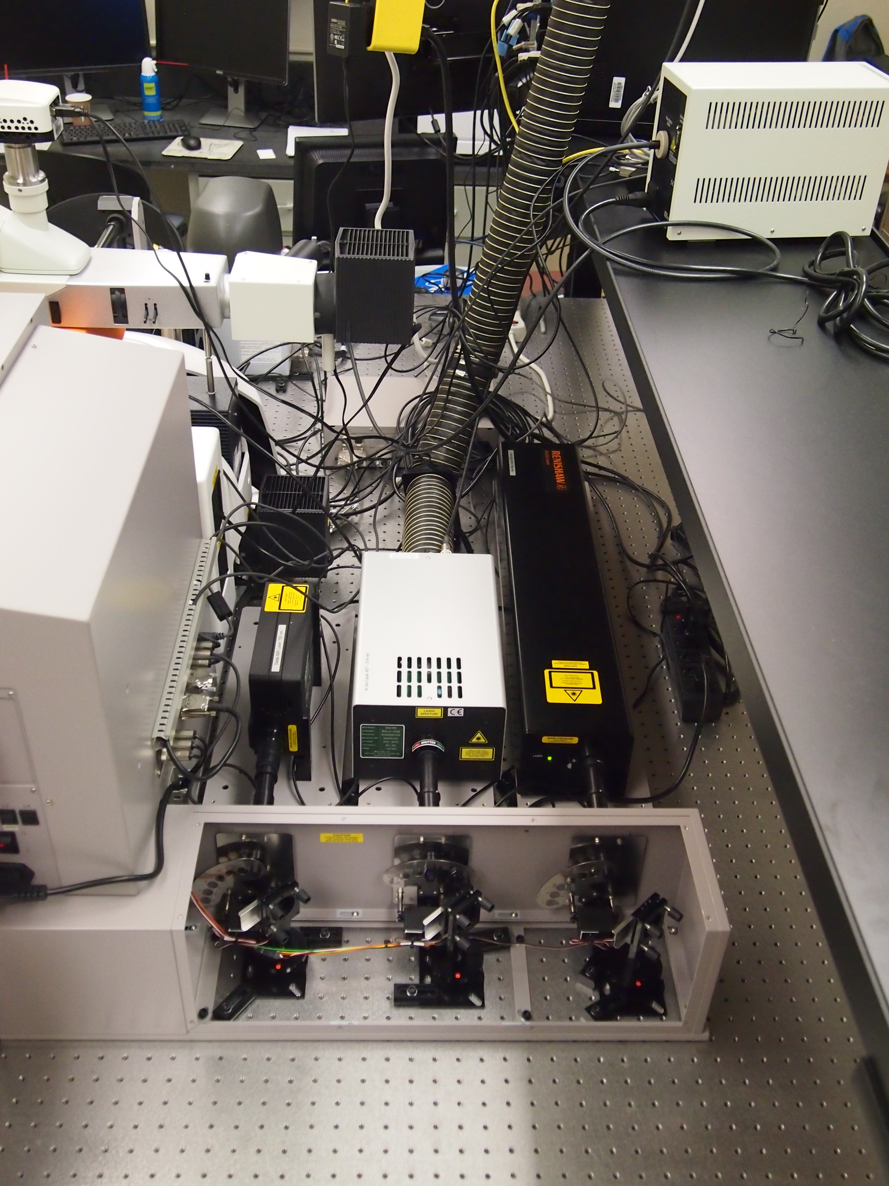
• 785 nm diode, 457/514 nm Ar ion and 633 nm He-Ne lasers (from left to right)
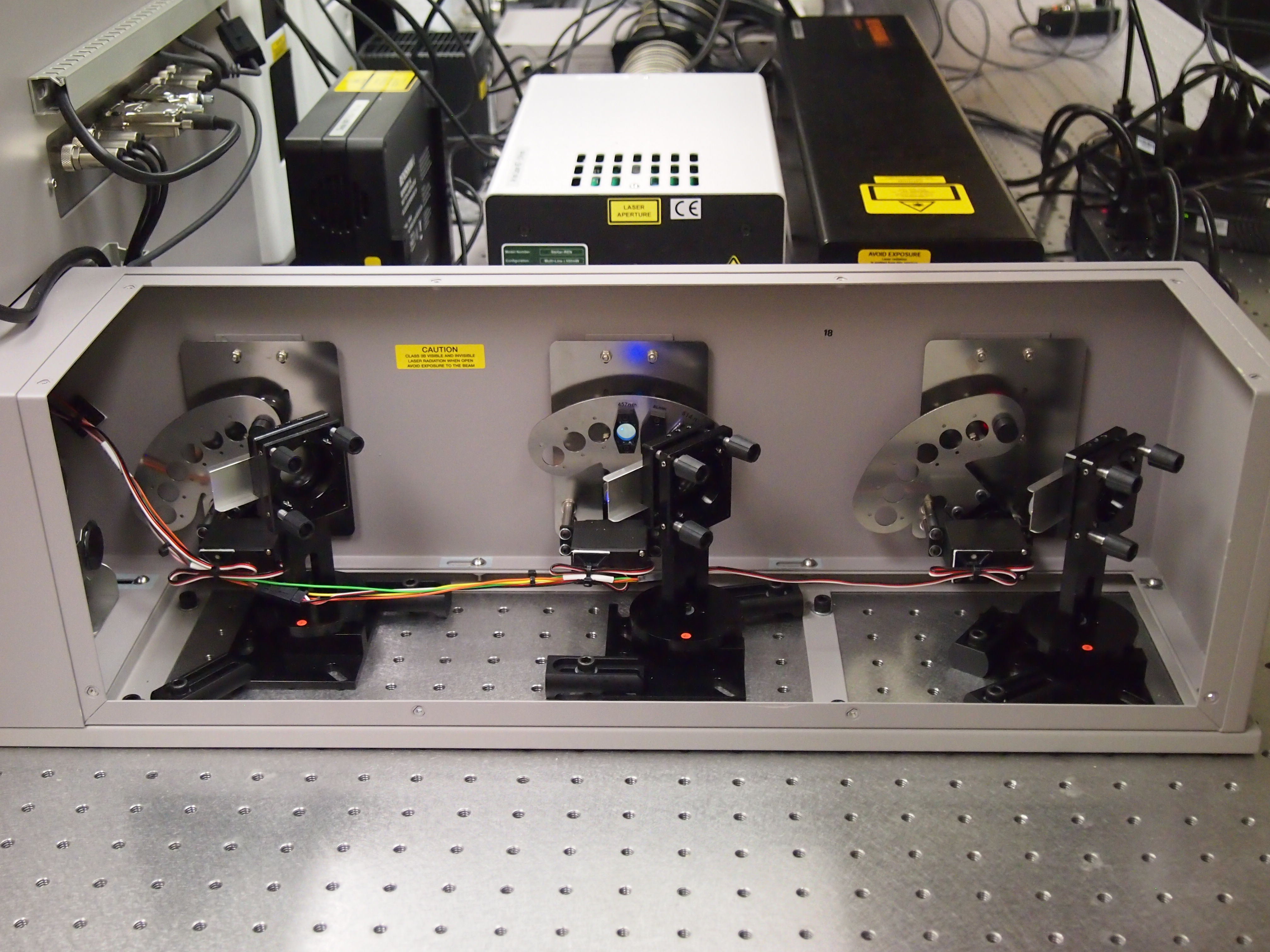
• Beamsteer motors and mirrors

• Spectrograph
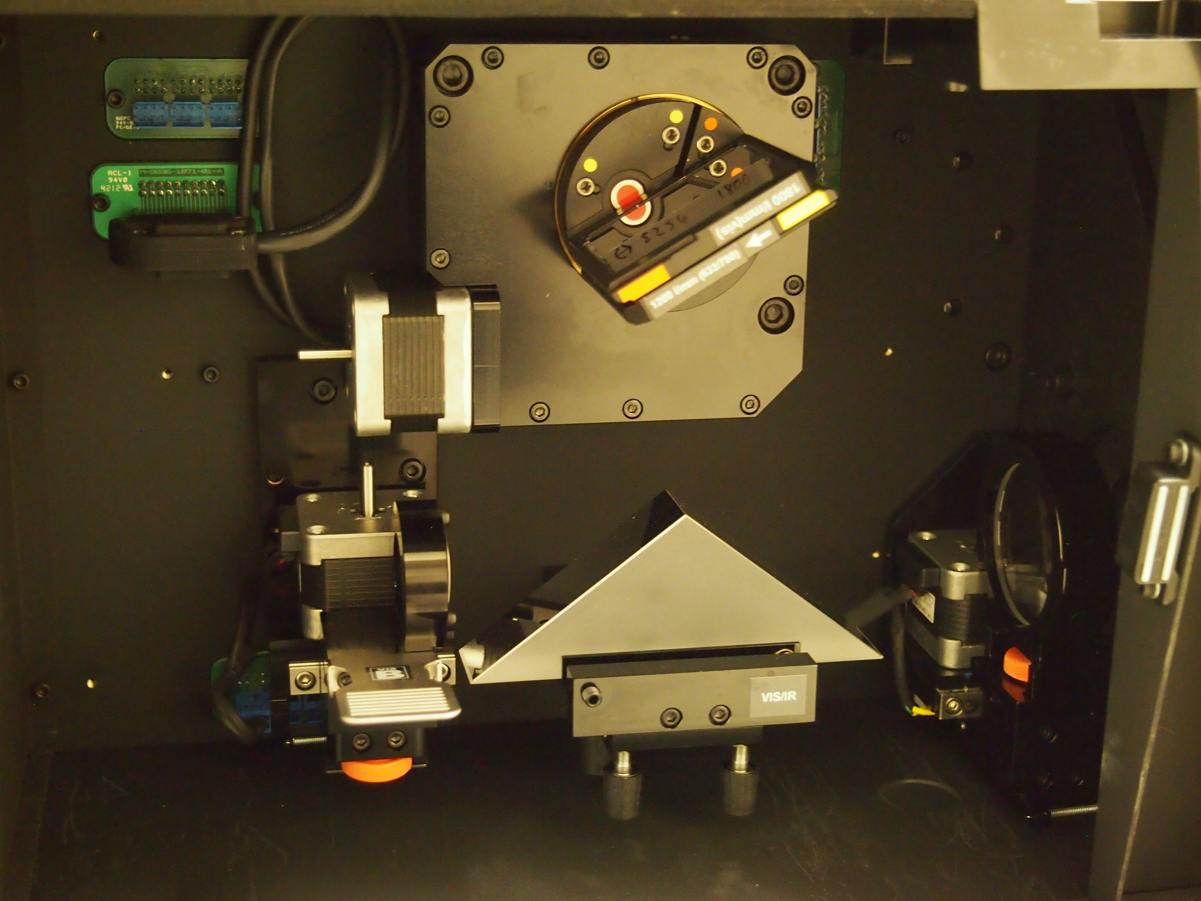
• Collimating lens, mirrors and focusing lens (below) and ultra-high precision spectrographic gratings (above)
Bruker Innova Atomic Force Microscope
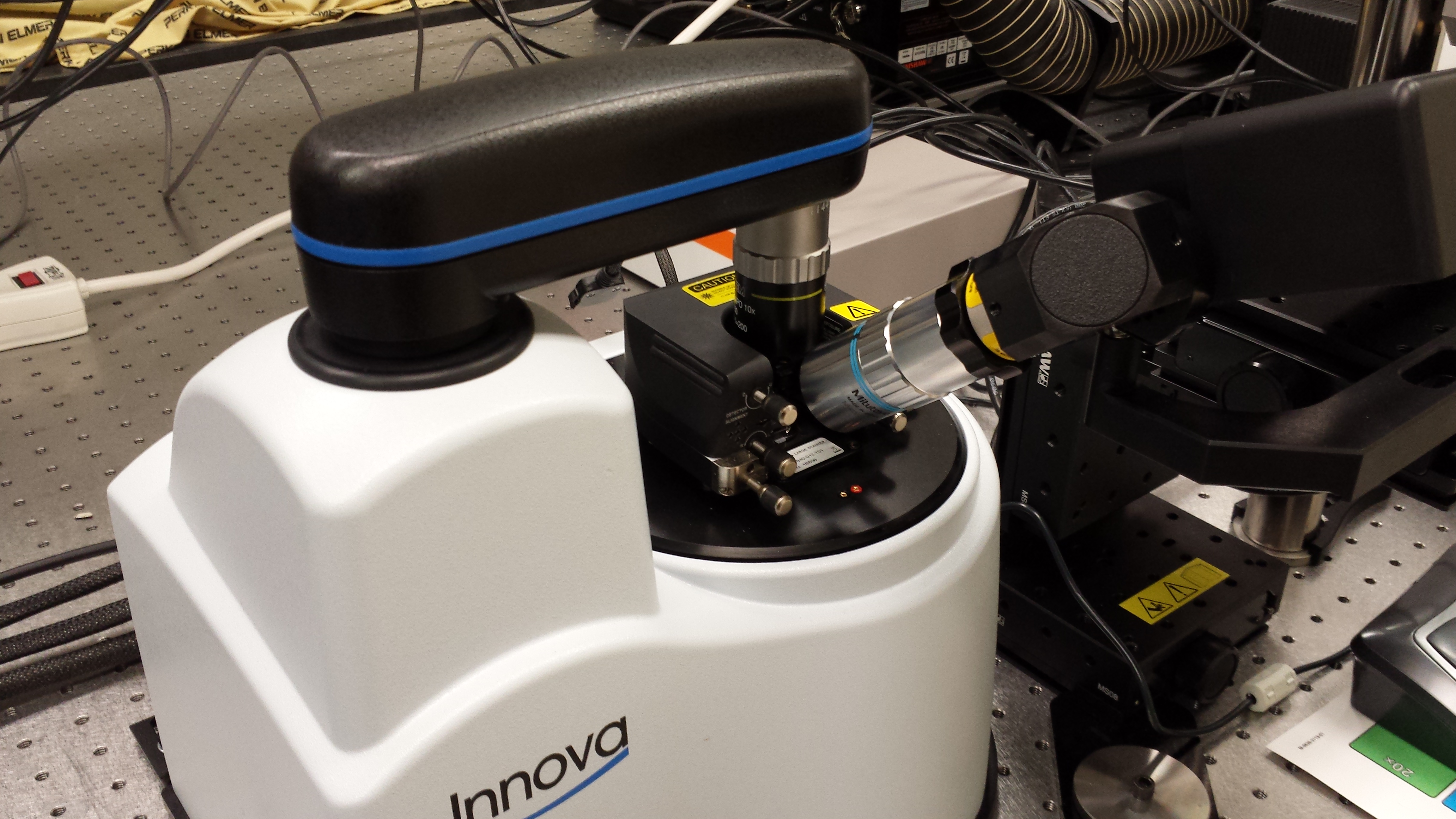
Bruker Innova Atomic Force Microscope includes:
• stacked piezo XY and Z scanners with resolutions as high as <0.02 and <0.01 nm, respectively
• high precision XYZ motorized stage
• optical AFM head for contact, tapping, lateral force, and phase imaging AFM and scanning tunneling microscopy (STM)
• optical AFM head for co-localization and tip-enhanced Raman scattering (TERS), improving Raman spatial resolution to <20 nm in XY and Raman scattering efficiency by >1,000-fold
• on-axis optical microscope with high-resolution (HR) video camera
• Trackball-guided motorized AFM optical coupler to Renishaw spectrometer that includes separate video optics, Raman interface IRIS software, a long working distance 60x objective for off-axis illumination of the AFM probe tip (~60° to probe axis) and collection of Raman scattered emissions.
• Special AFM-TERS package – tuning fork cartridge and Au-wire probes for non-conducting samples (biologicals and organic polymers)
• liquid MicroCell for aqueous AFM measurements
• NanoDrive control and NanoScope Image Analysis software

• AFM images (left to right: height, tapping amplitude and 3D topography) of dual-phase polymer film acquired in tapping mode
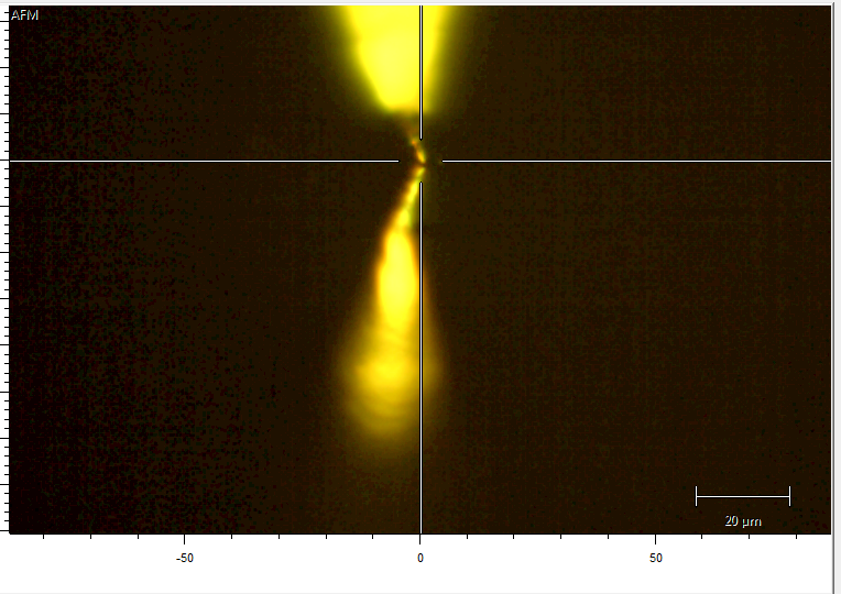
• Image of a gold TERS tip engaging the sample surface and its reflection obtained by video camera in Renishaw optical coupler to the AFM.
Linkam THMS600 Stage (-196° to 600°C)
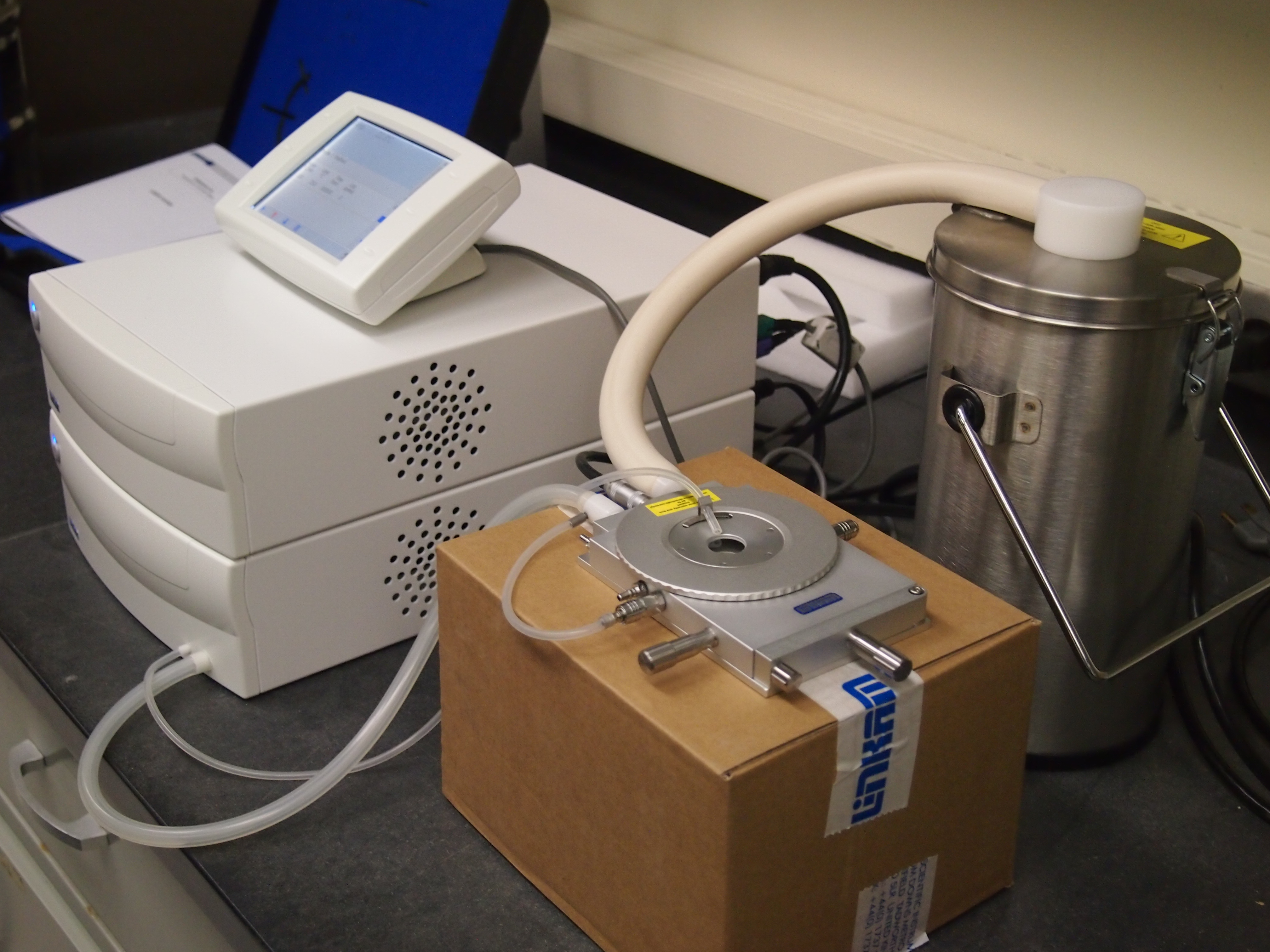
Linkam THMS600 Stage Specification:
• Temperature range -196° to 600°C
• Up to 150°C/min heating, 100°C/min cooling
• Temperature stability <0.1°C
• 16 mm X,Y sample manipulation
• Sample viewing area 22 mm diameter
• Quick-release gas valves for atmospheric control
• 100 ohm platinum resistor sensor. 1/10th Din Class A to 0.1°C
• Light aperture – 2.0 mm diameter
• Silver heating block for high thermal conductivity
• Direct injection of the coolant into heating block
• Single ultra thin lid sapphire window – 0.17 mm
• Objective lens working distance 4.5 mm
• Condenser lens minimum working distance 12.5 mm
• Range of condenser extension lenses available
• Can be used with all microscope techniques
• Water cooled stage body for high temperature work (>300°C)
• Suitable for Confocal, Laser Raman and X-Ray
• Sample side loading without removing the stage lid
Background Reading
What is Raman Scattering?
What is Atomic Force Microscopy?
What is Tip-enhanced Raman Scattering (TERS)?
